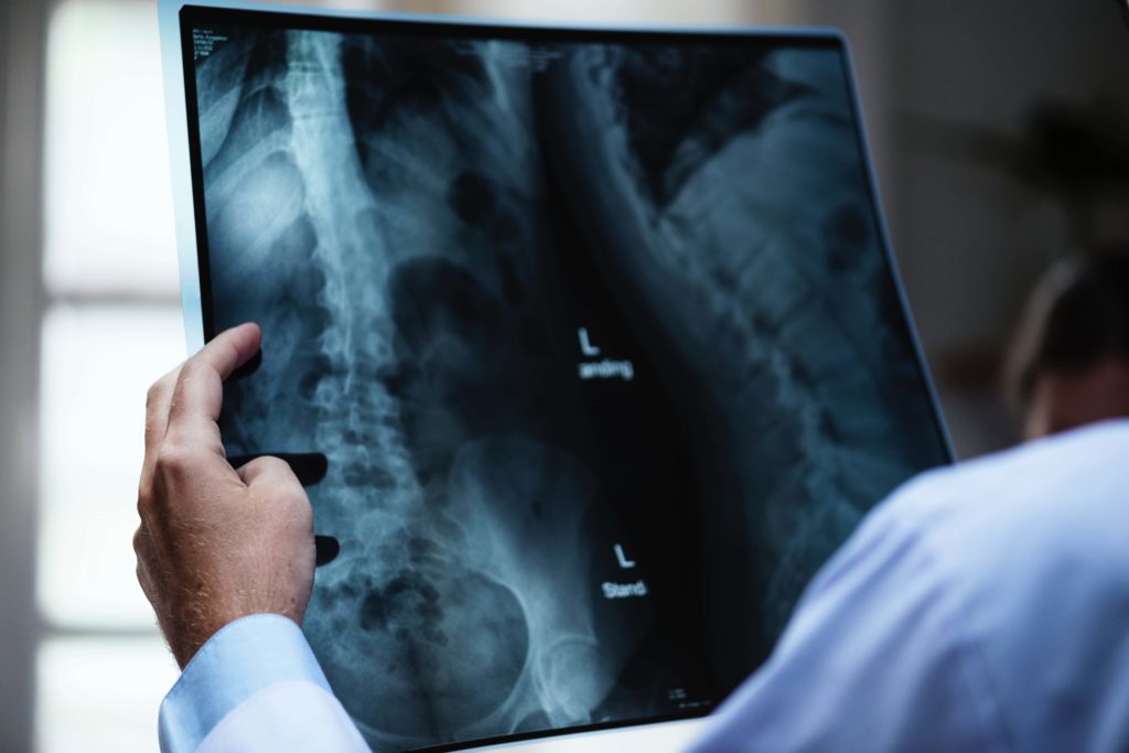
Radiology is the practice of reviewing images (including x-rays, MRIs and CT scans, among others) to diagnose illness or injury. Radiologists are specialized physicians who are an important part of a medical team and play a key role in the diagnostic chain.
Common errors include misdiagnosis/misreading an image, not doing the testing, failing to actually reporting what the image shows, and not following-up on testing.
A Radiologist’s Role on a Medical Care Team
As part of a routine checkup/screen, or in order to diagnose an illness, primary care physicians (family doctors) or physicians in hospitals will do assessments, obtain the history of a patient, and will order blood tests and other medical exams in order to diagnose what possible medical issues or conditions the patient may have.
The primary care physician may also order additional medical imaging studies to assist in this determination. Where that happens, the patient will have images taken by X-ray, CT, MRI etc and radiologist will review both these images as well as any background information and medical history and will given their impression of what the image shows and their opinion to help provide a diagnosis which will inform the direction of treatment for the patient. Accurate review and reading of these images is critical as this will form the basis for a patient’s diagnosis and treatment plan.
These images are taken in clinics or in-hospital by technicians. Once the images are ready, the radiologist reviews them and creates a formal report summarizing what they see on the image. They will send the report to the physician that ordered the imaging and to any other relevant medical personnel.
Radiology Techniques
X-Rays
Radiographic images (more commonly referred to as X-rays) are produced through a combination of ionizing radiation and light striking a photosensitive surface. X-rays produce images of the inside of a patient’s body, which show up in different shades of black and white depending on their ability to absorb the radiation. Bones will appear white (as calcium absorbs the most amount of rays), fat and other soft tissue will appear grey (as they absorb less rays), and other areas will appear black.
X-rays are often used to diagnose broken bones, but can be used to diagnose pneumonia (through chest x-rays) or breast cancers (through mammograms). It does not provide very detailed view and having too many X-rays can raise concerns about radiation.
CT Scans
Computed tomography (more commonly referred to as CT scans) are a series of specialized images that show doctors two dimensional (and now even three dimensional) “slices” of a patient’s body (much like you could look inside of a loaf of bread through slicing it). CT scans are non-invasive and can be performed relatively quickly both at hospitals and outpatient clinics.
CT scans have allowed physicians to see diseases and problems that, in the past, could only have been seen through performing surgery. They can help doctors see small nodules or tumours that do not appear on a conventional film x-ray. They are often used to evaluate the brain, sinuses, neck, chest, spine, abdomen and pelvis.
Like x-rays, CT scans utilize radiation to produce the images and should therefore be only used when necessary.
MRIs
Magnetic resonance imaging (more commonly referred to as an MRI) is similar to a CT scan in that it also produces cross-sectional images of a patient’s body. Unlike a CT scan, an MRI does not use radiation to produce the images. Instead, it uses a strong magnetic field in combination with radio waves to create detailed images of the inside of a patient.
MRIs are commonly used to examine the brain, spine, abdomen, pelvis, and joints. A specialized form of MRI, known as magnetic resonance angiography (or MRA) can be used to examine blood vessels. This is the most sensitive test and is especially important when diagnosing and treating brain injuries due to hypoxia, trauma, or other conditions.
MRIs can be used alone or to further evaluate an abnormality captured by a CT scan.
Ultrasounds
Ultrasonography, also known as ultrasound, is a medical imaging technique that send pulses of ultrasound into tissue through a probe, and using the echoes of ultrasound pulses to delineate objects or areas of different density in a patient’s body in order to produce images. Ultrasound pulses are sound waves with frequencies that are much higher than those that are audible by humans.
In addition to their popular use in imaging fetuses of pregnant women (also known as obstetric ultrasound), ultrasound can also be used to see internal body structures such as organs, tendons, muscles, joints and blood vessels.
Ultrasounds are less sensitive than MRIs, but they are still very useful in determining whether or not a baby has sustained a brain injury around the time of birth (neonatal encephalopathy). However the timing of the ultrasound is critical- if the ultrasound is done too early or too late, it will miss the injury.
What is fascinating about ultrasounds is that the they are able to see the brain of a baby, but not of an adult. The adult skull is too thick to allow the ultrasound to work properly.
No radiation is used in this type of imaging, and it is particularly useful to image pregnant women or children.
PET Scans
A positron emission tomography scan (also known as a PET scan) is an imaging test that uses a special dye with radioactive tracers that is swallowed, inhaled, or injected depending on which part of a patient’s body is being examined. Once the tracers are administered, the patient is scanned with a gamma camera or a positron emission scanner. After being absorbed by the patient’s organs and tissues, the tracer will collect in areas of higher chemical activity (associated with disease), which will show up as bright spots on the PET scan.
PET scans are used to diagnose cancer, heart problems, issues with the central nervous system, and brain disorders.
Common Radiology Errors
- Failing to do the testing:
Obviously, the imaging study was ordered for a reason- because it was important to the timely diagnosis and treatment of the patient. If the radiologist (or the radiology clinic/department) fails to do the imaging study, it can lead to serious consequences for the patient.
- Failing to report the results:
If a radiologist’s report is not actually sent/received by the physician who ordered it in a timely way, it can cause significant delays in diagnosing and treating the patient. Similarly, if the ordering physician receives the report but doesn’t actually review it (for example, if it gets lost or sits in their inbox), this may constitute negligence.
- Failure to follow-up on results:
This is similar to the issue of failing to report the results. If a serious problem or condition is detected by the radiologist, or if the testing is unclear, they have an obligation to repeat the testing or recommend further testing. They have an obligation to report this to the ordering physician.
- Diagnostic Errors:
Some common examples of radiology errors include serious diagnostic errors such as:
- Misreading a spinal cord MRI (especially when there is stenosis);
- Failure to see a pulmonary embolism;
- Missing a major infection or abscess;
- Mistaking cardiopulmonary failure for some other illness;
- Mistaking a cancerous tumour for a benign one;
- Failure to see a bowel obstruction;
- Failure to see an aneurysm;
- Misdiagnosis of brain injury;
- Misreading mammogram findings.
Radiology Errors and Medical Malpractice
If you believe you were misdiagnosed by a radiologist or there was a delay in diagnosis, you may have grounds for a medical malpractice claim against the radiologist and/or your primary care physician.
At Sommers Roth & Elmaleh we have been representing patients affected by misdiagnosis and failure to diagnose for many years. In some cases, clients may not have even realized that ongoing symptoms they are experiencing or medical complications they are dealing with may be a result of a misdiagnosis. We work with medical professionals and other experts to carefully review your medical records to establish error or negligence on the part of a physician or other health care worker. We also provide trusted and compassionate guidance to our clients throughout the process of pursuing their claim.
If you have questions about the medical care you received, or if you suspect that you were misdiagnosed or incorrectly diagnosed, contact the respected and highly knowledgeable Toronto medical malpractice lawyers at Sommers Roth & Elmaleh in Toronto. We have been involved in many precedent setting decisions and have a proven track record of success. Call us at 1-844-777-7372 or contact us online for a free consultation.

